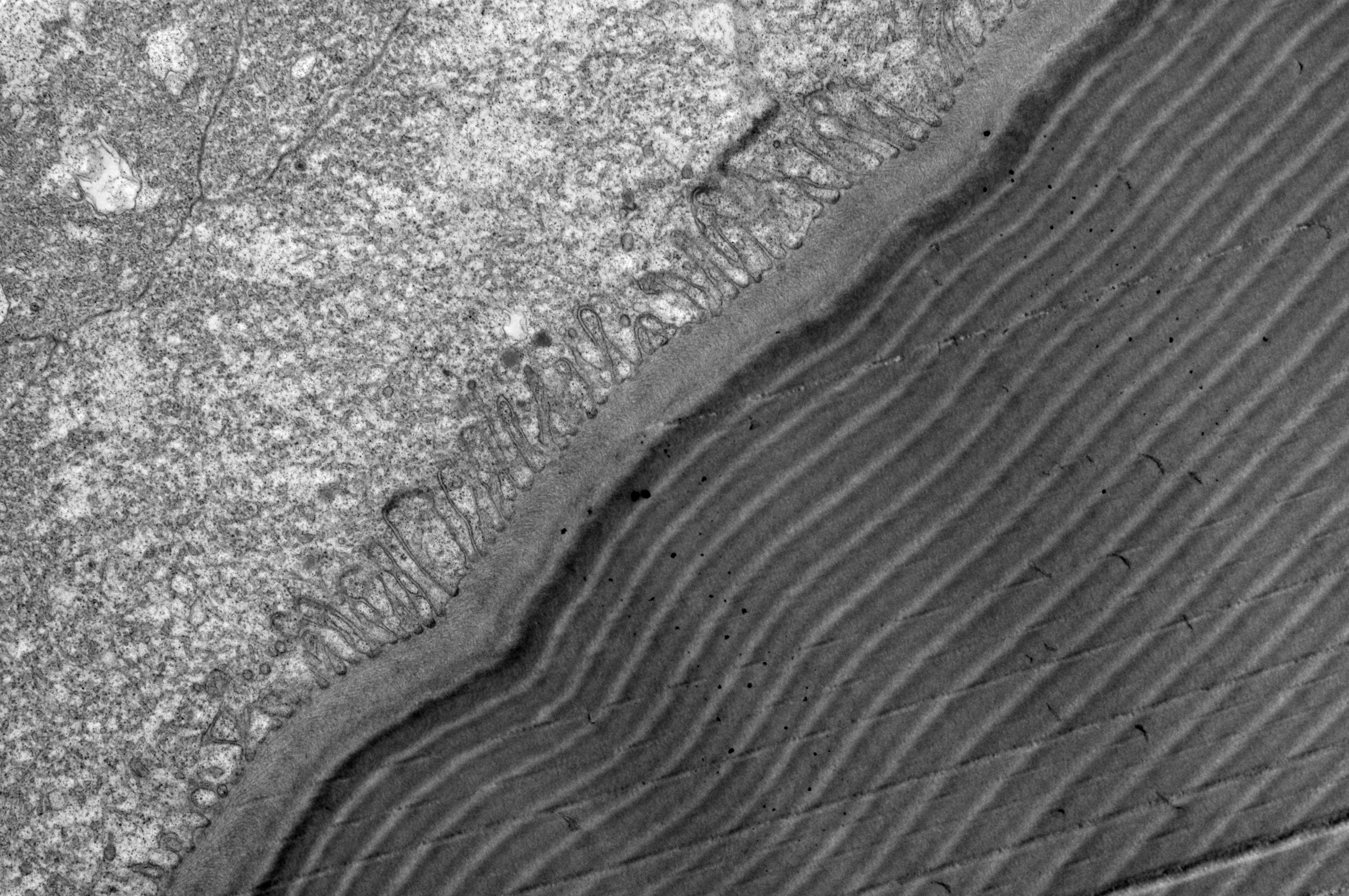
Sunburst diving beetle (Thermonectus marmoratus) larval lens imaged using transmission electron microscopy. Lightly stained chitin fibrils are arranged in a darkly stained protein matrix
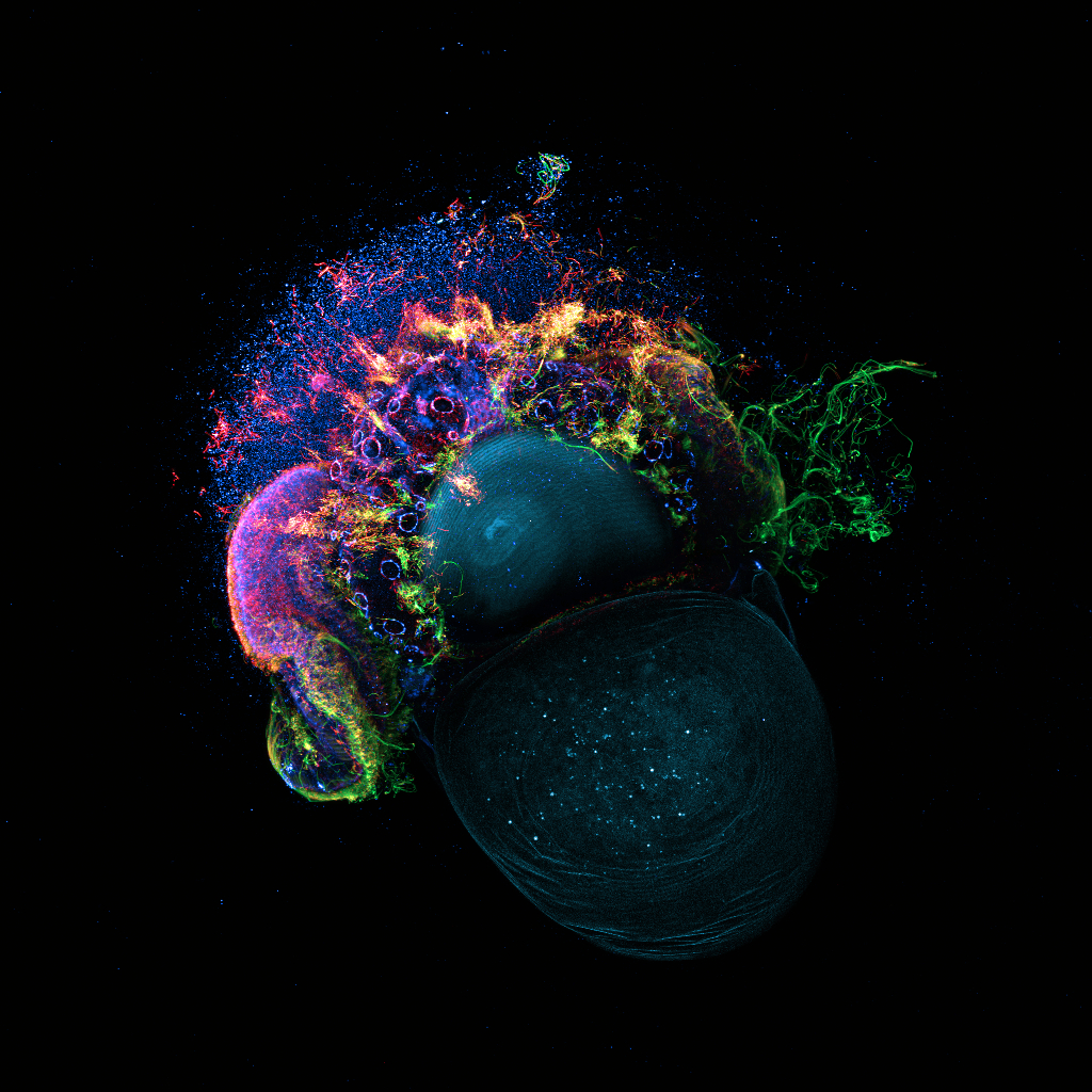
Slipper limpet (Crepidula fornicata) veliger larva imaged using confocal microscopy. Stained with DAPI and antibodies against acetylated tubulin, ⍺-tubulin and 𝝱-catenin in cyan, red, green and blue respectively.
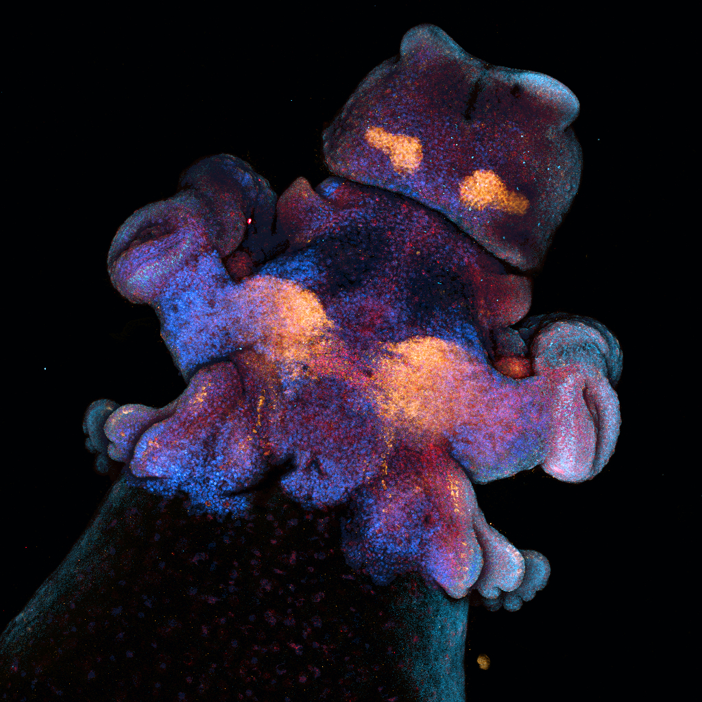
Longfin inshore squid (Doryteuthis pealei) embryo. Nuclei and mRNAs for elav, optix, and pcdh17 mRNAs represented in blue, red, cyan and orange respectively. Imaged using confocal microscopy.
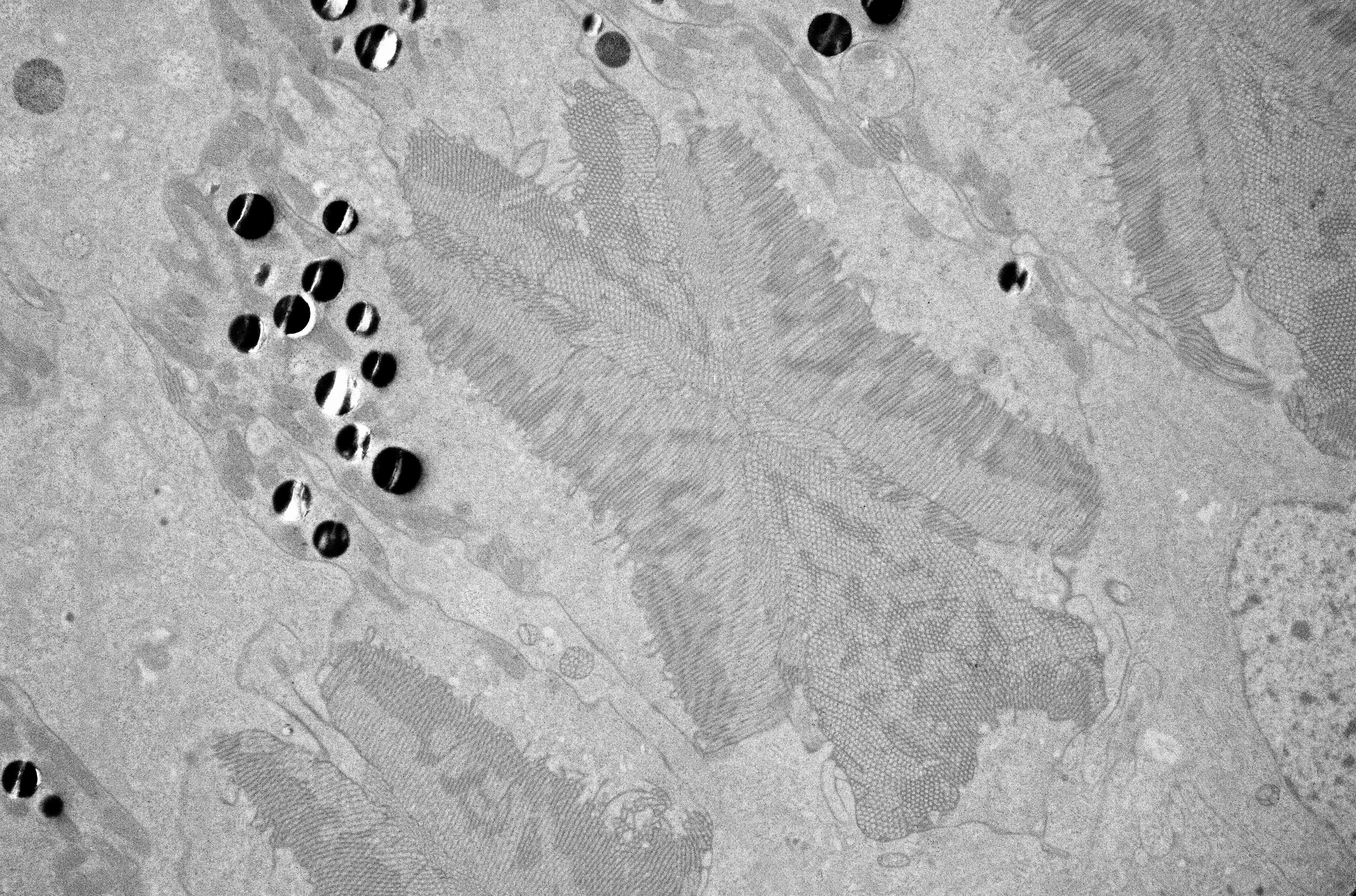
Redhead pine sawfly (Neodiprion lecontei) larval photoreceptors. Imaged using transmission electron microscopy.
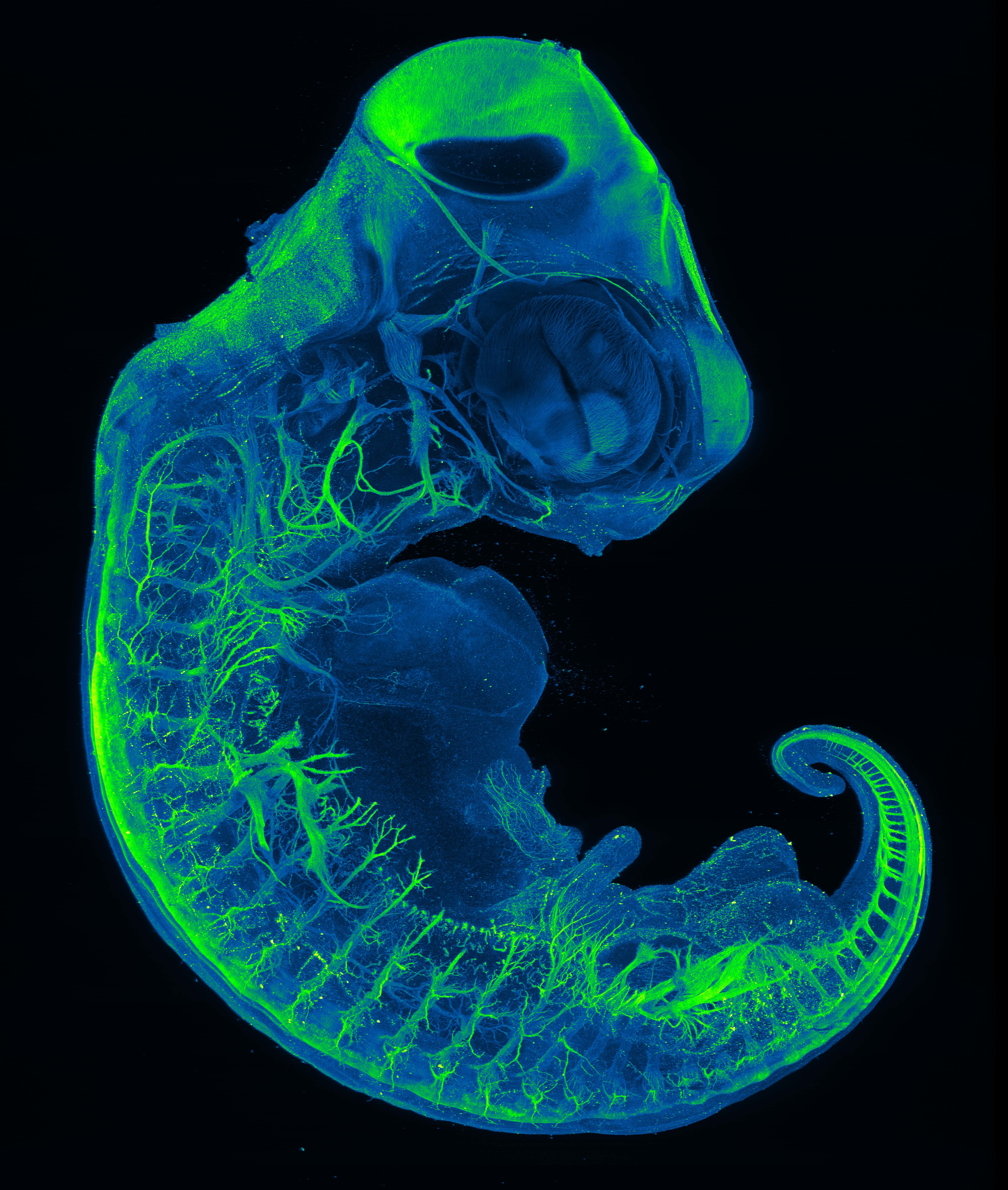
Red-eared slider (Trachemys scripta) embryo stained with acetyl-alpha tubulin, labelling neurons. Optically cleared and imaged using confocal light sheet microscopy.
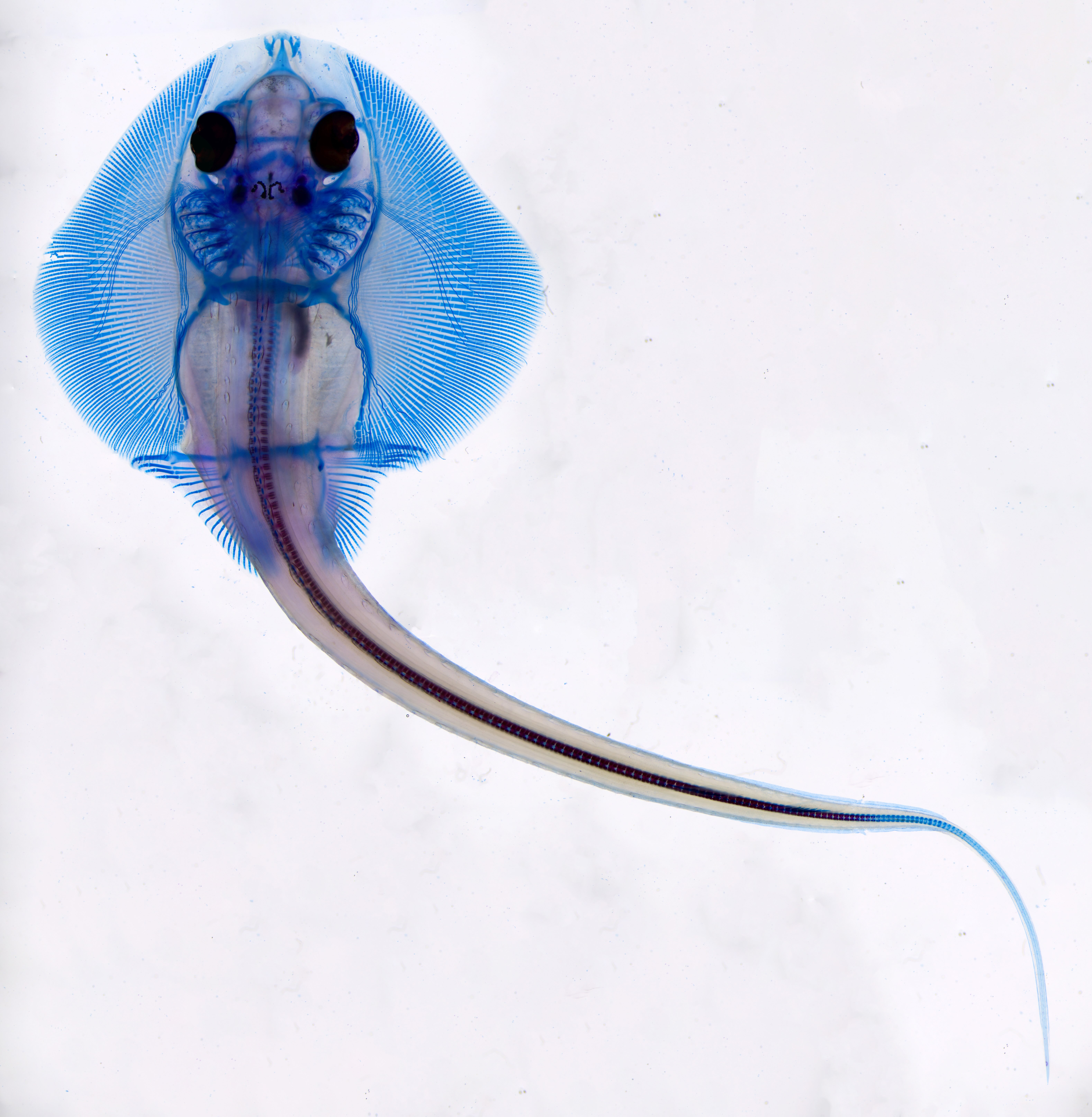
Skeletal preparation of little skate (Leucoraja erinacea) embryo labelling cartilaginous tissue in blue and calcified tissue in red. Assembled using focus stacking and tile stitching.
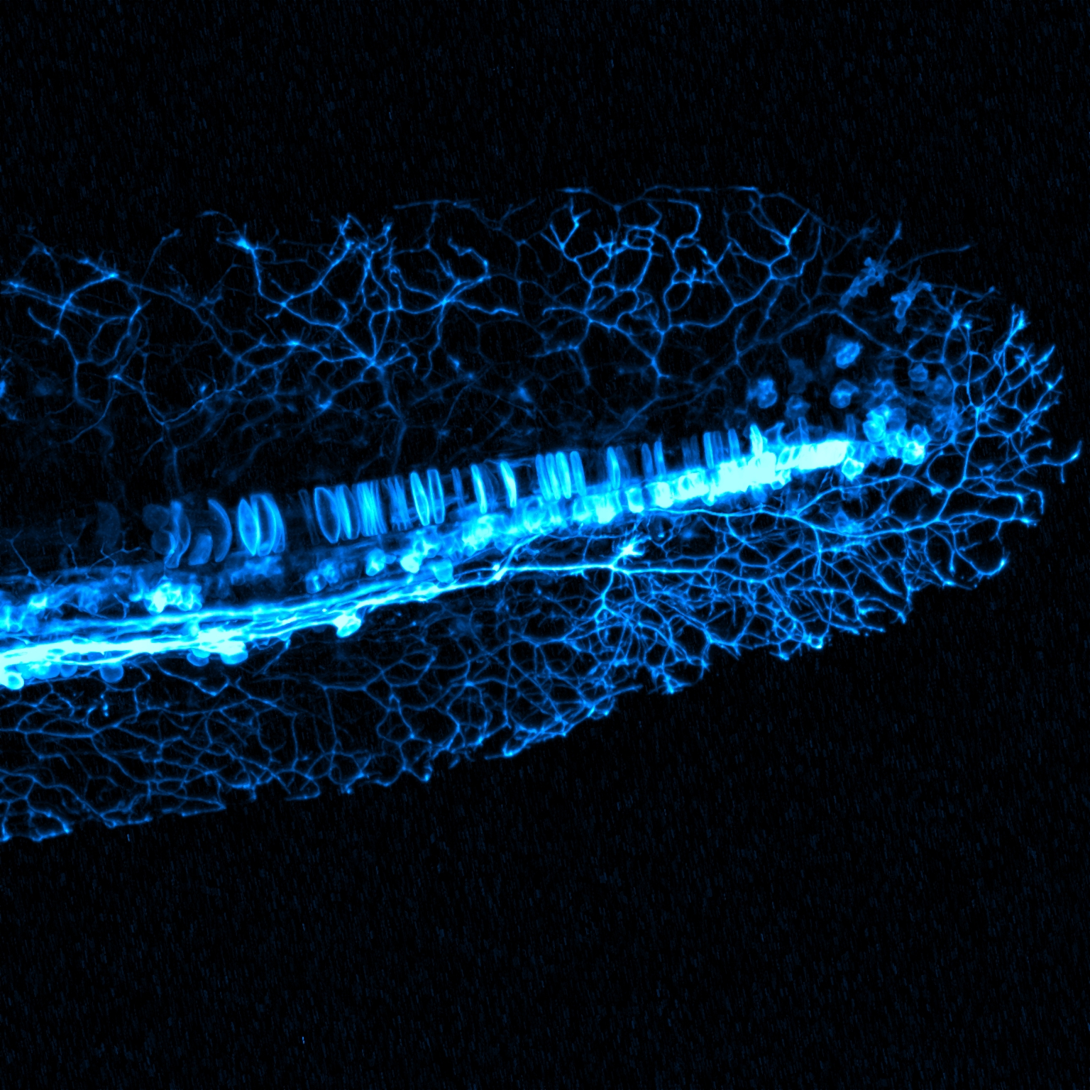
Differentiating neurons in a transgenic (GFP tagged neurogenin) embryonic zebrafish (Danio rerio) tail
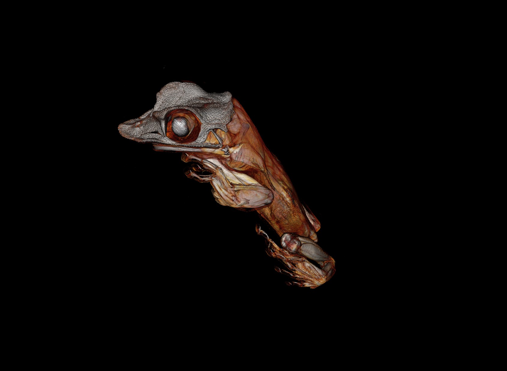
Artifcially colourised 3D rendering of Yucatán casque-headed tree frog (Triprion petasatus). Captured with Micro-CT